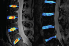
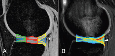
In close collaboration with the clinical musculoskeletal section, MQIR aims to integrate research and build collaborations between basic scientists, clinical scientists, and physicians, establishing a strong resource for musculoskeletal imaging-based research at UCSF. Recognizing that collaborative bridges between departments and campuses is key to advancement in today's research climate, MQIR strengthens and nurtures partnership not only within the Department of Radiology, but also between the Departments of Orthopaedic Surgery, Medicine and Bioengineering at UCSF and UC Berkeley.
Central to the MQIR research effort is a Bioengineering Research Parntership grant (Principal Investigator: Dr. Sharmila Majumdar) from the National Institutes of Health, which has led to collaborations with Dr. Jeffrey Lotz, Dr. Michael Ries, Dr. Marc Safran, and Dr. Ben Ma from Orthopaedic Surgery, Dr. Thomas Budinger from Bioengineering, and UC Berkeley. The BRP team focuses on developing state-of-the-art quantitative imaging techniques for studying cartilage (particularly in the knee) as well as intervertebral discs in the spine.
In addition, Dr. Majumdar's research utilizes micro-computed tomography, infrared spectroscopic imaging and magnetic resonance imaging to quantify microscopic bone structure in osteoporosis and osteoarthritis. In an effort to push the realm of musculoskeletal imaging to include high resolution, morphological and molecular imaging, her research involves extensive collaborations with Dr. John Kurhanwicz, Dr. Sarah Nelson and Dr. Dan Vigneron, all in Radiology. Dr. Majumdar's group includes postdoctoral fellows, researchers, graduate students, medical students, visiting scholars, and residents.
MQIR has access to multiple tools for imaging and analysis in musculoskeletal research.
We hypothesize that high resolution imaging techniques combined with quantitative image analysis, processing and visualization can provide new insights and clinically viable and relevant methods for objective evaluation of disorders of the musculoskeletal system. The long-term objective of our research is to understand the link between morphology, function and clinical symptoms in the musculoskeletal system. Therefore, we facilitate close interactions between bioengineers, computer scientists, clinical investigators, basic scientists, and corporate partners. This effort will expedite the development of clinically relevant quantitative imaging tools and propel the technical advances from laboratories into operating rooms and clinics.
Research directions
High field and high resolution magnetic resonance imaging for quantitative characterization of the morphology and function of the musculoskeletal system, with particular attention to bone, cartilage and intervertebral disc and diseases such as osteoporosis, spinal disorders and osteoarthritis. Microscopic characterization of bone, cartilage, disc, and other tissues using methodologies such as computed tomography, Fourier Transform Infrared Spectroscopy, high resolution nuclear magnetic resonance spectroscopy, and confocal laser microscopy. Development of high resolution and quantitative computed tomography for characterizing bone geometry, micro-architecture and density, aimed at understanding aging, ethnic differences in the skeleton, osteoporosis, and orthopedic implants.