Commentary on Breakthrough COVID-19 Infections from Brett Elicker, MD

Breakthrough viral infection - when a fully vaccinated person gets infected with COVID-19 – seem to be more common with the fast-spreading Omicron variant. Recently Brett Elicker, MD, chief of Cardiac & Pulmonary Imaging Section in the UC San Francisco Department of Radiology and Biomedical Imaging, provided commentary on these breakthrough infections, what they are and what they look like.
“Breakthrough infections after COVID-19 vaccination are uncommon and typically present with mild symptoms and radiologic abnormalities that resemble primary COVID-19 infection, albeit less severe,” he says. “As the pandemic continues and the length between vaccine administration lengthens, it is likely that the severity of breakthrough infections will increase and thus radiologists should be aware of the possibility of breakthrough infections and their typical imaging findings.”
Dr. Elicker’s full commentary was recently published in Radiology: Cardiothoracic Imaging where he is associate editor. The commentary was in relation to a published retrospective study conducted by radiologists at the Department of Radiology, University of Maryland School of Medicine which characterized chest radiographic and CT imaging appearance in patients with breakthrough COVID-19 in a hospital setting.
Through studies, we can determine that breakthrough respiratory viral infections can occur for a variety of reasons including:
- Inadequate antibody response resulting from an immunocompromised state
- Individual's immunity waning over time
- New strains of the virus showing resistance to previous immunizations
 As a specialist in thoracic radiology and a particular interest in evaluating interstitial lung disease, Dr. Elicker notes that the pulmonary manifestations of viral infection represent a combination of direct injury by the viral organisms and the immune response to their presence. However, the immune response is likely a more important driver in the moderate to severe forms of lung injury.
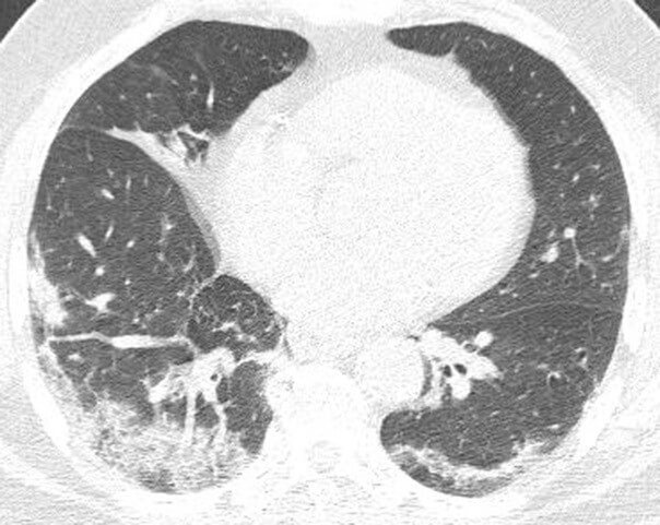
During the COVID-19 pandemic, radiologists continue to study chest radiographic and CT findings of patients with COVID-19 to determine patterns. Common imaging findings of nonbreakthrough (or typical) COVID-19 cases include bilateral consolidation and ground-glass opacity that may be peripheral or diffuse in distribution. According to Dr. Elicker, the imaging appearance suggests that organizing pneumonia is a relatively common pattern of injury in these patients as evidenced by the presence of perilobular opacities and/or the reversed halo sign in many patients. More severe manifestations include diffuse alveolar damage, which on imaging manifests as generalized ground-glass opacity and consolidation. “It is known that both diffuse alveolar damage and organizing pneumonia may, over time, result in lung fibrosis,” he says.
Findings from the University of Maryland study, led by Rydhwana Hossain, MD show that imaging findings in such patients are commonly mild and outcomes are generally favorable. The authors note that “larger studies are needed to establish imaging differences between breakthrough and unvaccinated populations.”
Overall, as the number of breakthrough COVID-19 cases increase, it’s important as radiologists to document imaging findings to determine if the imaging patterns remain consistent with those previously observed. It’s also noted that larger studies are needed to establish imaging differences between breakthrough and unvaccinated populations.
