Exploring the Brain: How Are Brain Images Made with CT?
The following article was written by Christopher P. Hess, M.D., Ph.D, and Derk Purcell, M.D, Assistant Clinical Professor in the Department of Radiology and Biomedical Imaging at UCSF.
The complexity of the organ that determines how a person thinks, moves, feels, and remembers is overshadowed only by its unique vulnerability. The brain is hidden from direct view by the skull, which not only shields it from injury but also hinders the study of its function in both health and disease. The cells in the arteries that supply the brain are so tightly bound that even most normal cells in the bloodstream are prevented from crossing the so-called “blood-brain barrier,” thereby rendering the normal chemistry of the brain invisible to the routine laboratory blood tests that are often used to evaluate the heart, liver or kidneys.
Computed tomography (CT) and magnetic resonance imaging (MRI) have revolutionized the study of the brain by allowing doctors and researchers to look at the brain noninvasively. These diagnostic imaging techniques have allowed for the first time the noninvasive evaluation of brain structure, allowing doctors to infer causes of abnormal function due to different diseases.
____________________
Computed tomography is based on the measurement of the amount of energy that the head absorbs as a beam of radiation passes through it from a source to a detector. Within a CT scanner, the radiation source and detector are mounted opposite one another along a circular track, or gantry, allowing them to rotate rapidly and synchronously around the table on which the patient lies. As the x-ray source and detector move around the patient’s head, measurements consisting of many projections through the head are obtained at prescribed angles and stored on a computer. The table moves horizontally in and out of the scanner in order to cover the entire head.
The “tome” in tomography is the Greek word for “slice.” At the core of the scanner is a computer that not only controls the radiation source, the rotation of the x-ray tube and detector, and the movement of the table, but also generates anatomical slices, or tomograms, from the measured projections.
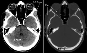
The primary physical quantity that is captured with CT is density, or mass per unit volume. Prior to display and storage of CT images, pixel intensities are mapped to a standard numerical scale to allow reliable discrimination between different densities of tissue such as air, water, fat, bone, and various brain constituents. When the images are reviewed on a computer, the intensities are further modified by a process referred to as windowing in order to optimally depict the density of different tissues for visual display. Extremely dense material, such as metal or bone, appears bright on CT images, whereas tissue that is less dense, like fat or water, appears dark.
At this point, the interpretation of brain images requires a detailed knowledge of anatomy and a comprehensive understanding of how different diseases affect the brain and its supporting structures. Radiologists are medical doctors who specialize in acquiring and interpreting images, while neuroradiologists focus specifically on imaging of the nervous system. These specialists work together with neurologists, neurosurgeons and primary care physicians to use CT and MRI to diagnose disorders of the brain and understand their significance for patients.

