Risk Reduction at UCSF

Risk Reduction at UCSF

At UCSF we are very concerned about radiation—after all, we work around it every day.

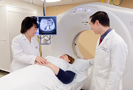
The Department of Radiology and Biomedical Imaging is fully committed to minimizing exposure to radiation for our patients and staff. We follow the principle of "ALARA" (radiation dose As Low As Reasonably Achievable) when it comes to radiation and patient safety. That means we always use the lowest radiation dose consistent with getting imaging results that are of appropriate diagnostic quality.

Low-dose CT Protocols at UCSF Medical Center

UCSF uses CT settings (protocols) that adjust the dose of radiation depending on the patient size, the body part being imaged, and the reason for the study.  These protocols are routinely reviewed for each body part.  Specific protocols are used for pediatric imaging, which minimize dose.

UCSF Ensures Imaging Safety

UCSF’s Department of Radiology & Biomedical Imaging is fully committed to minimizing exposure to radiation for our patients and staff. Here are things we routinely do to ensure imaging is safe:

  1. Choosing the most appropriate imaging study:  Minimizing the exposure to radiation dose begins with the proper selection of an imaging study for the specific needs of every individual patient. Our radiologists are available 24 hours a day, seven days a week to consult with referring doctors regarding the relative benefits of tests that may or may not involve radiation exposure, and we encourage referring physicians to consider tests that do not involve radiation (e.g., ultrasound or MRI) if they can provide the same information as a CT scan or other test with radiation. Specific examples include using MRI instead of CT to evaluate inflammatory bowel disease and ultrasound to diagnose appendicitis in pregnancy.
  2. Tailored techniques:  At UCSF, specific radiation dose protocols are in use and routinely reviewed for each body part, and special low radiation dose protocols have been developed for certain indications in pediatric patients and during CT-guided procedures.
  3. Careful quality control:  To further minimize the potential risks of radiation, quality control procedures are performed on a scheduled basis to assure that all X-ray imaging devices are operating correctly and safely, and all machines within the Department of Radiology & Biomedical Imaging are tested at least annually. Also, quarterly maintenance is performed on all CT scanners in the Radiology Department to ensure that they are operating to specification. We are in compliance with all national standards for radiation safety.
    In addition, radiation levels for CT and Interventional Radiology procedures are monitored. At the end of each study, dose measures are sent to and stored in a separate database.  Values for each procedure are compared to those stored in the database, and threshold levels have been set; if those levels are exceeded, it will cause an immediate warning e-mail to be sent to the appropriate staff responsible for overseeing and following up on these exposures.
  4. Latest CT technology for automatic radiation dose reduction:  All UCSF CT Scanners are equipped with the latest generation automatic dose modulation software that reduces the radiation dose to various body parts during a scan. This software tailors the amount of radiation output depending on the body part being imaged and the size of the individual. For example, less radiation is needed to image parts of the thorax than the abdomen, and less is needed for a smaller person or smaller body part than a larger one. Dose in some areas of the body can be reduced by as much as 50 percent compared to CT scanners lacking this technology. UCSF has also installed on all its CT scanners image reconstruction software that further reduces dose by up to 30 percent while providing images with the same diagnostic information. We continually invest in dose reducing technology as it becomes available.
  5. Special attention to pediatric patients:  At UCSF we are very concerned about radiation exposure to our pediatric population.  Several of our radiologists specialize in infant imaging. When possible, we often recommend alternative imaging studies with ultrasound, which require no radiation at all.
    When it is necessary to image infants, we perform the imaging with portable, state-of-the-art radiographic equipment. We use collimation or coning, techniques that restrict the X-ray beam so only the region of interest is emphasized. The X-ray output is adjusted according to the infant’s size. Since premature infants are extremely small, the amount of radiation used for such radiographs is a tiny fraction of what is needed for adult imaging or CT. When a CT scan is necessary, we have special low radiation dose protocols for children.
    Radiologists who specialize in pediatric imaging interpret the images.
  6. New low-dose CT protocols:  In addition to protocols mentioned elsewhere, UCSF follows other new low-dose protocols for imaging, which include CT-guided spine procedures,  low-dose CT for early detection of lung cancer, and low-dose coronary CT angiography
  7. Beam collimation policy:  For general X-ray procedures, the X-ray beam is carefully restricted to the area of clinical interest, a process called collimating. 
  8. Training of technologists and radiologists:  Ongoing education of all technologists and doctors in our department is a key to maintaining radiation safety and the practice of “ALARA” (radiation dose As Low As Reasonably Achievable). In addition, training of personnel and the use of low-dose methods were integral factors in our CT scanners receiving accreditation from the American College of Radiology.
  9. Radiation Oversight Committee:  The UCSF Department of Radiology & Biomedical Imaging maintains a Radiation Oversight Committee that oversees radiation-intense protocols and education regarding imaging algorithms to minimize exposure to radiation when possible, and specifically:
  • Reviews and monitors CT radiation doses administered across all departmental sites.
  • Formulates and implements dose reduction strategies (both with respect to modality utilization patterns and technical protocols), emphasizing the department subspecialties with high volumes where dose reduction would have the greatest impact (i.e., Abdominal Imaging, Thoracic Imaging, Neuroradiology, Pediatric Radiology, and Nuclear Medicine).
  • Reviews and enhances educational tools and materials for physicians (both inside and outside the department) with respect to appropriate radiation utilization and radiation dose magnitude risks.
  • Considers targeting certain referring physicians or groups for specific practice modifications, and develops audit tools to monitor and document such successes.
  • Reviews and enhances radiation dose educational materials for patients.

CT-guided spine procedures

UCSF radiologists use many techniques, including CT-guided injections, to accurately diagnose back pain and tailor a personalized treatment. CT-guided injections allow more accurate placement of needles for pain treatment than conventional X-ray imaging used at most imaging centers.

The UCSF Imaging Center at China Basin is one of the few facilities in Northern California to use special low-dose CT "step and shoot" mechanisms during CT-guided spine procedures.  By using the latest dose reduction software and by changing procedural techniques, we have been able to reduce the total radiation dose to patients during CT-guided injections by 80 percent. This dose reduction allows procedures to be performed with the same or even smaller radiation effective doses than conventional X-ray fluoroscopy.  Precise, but low-dose guidance for needle placement in spine interventions allows delivery of medication to sites where pain is generated in the back, including disc herniations, facet joints and compressed nerves.  Learn more

Low-dose CT for early detection of lung cancer

A recent study from the National Lung Screening Trial showed a 20 percent reduction in lung cancer-related deaths in heavy smokers screened annually with low-dose CT compared to standard chest X-rays. Using low-dose CT, we are able to find cancers when they are much smaller and more treatable, as compared to standard chest X-rays, which only detect larger masses.  Learn more

Low-dose coronary CT angiography

We are using low-dose coronary CT angiography (CTA) to image the coronary vessels with substantially less radiation than traditional nuclear stress testing in selected patients. Prospective electrocardiographic gating techniques, which can be used in patients with lower heart rates, activate the CT scanner only at precise times during the cardiac cycle.

Risk Reduction at UCSF