CT-Guided Interventional Spine

Minimally invasive techniques are used to diagnose and treat vertebral disc problems and many other conditions of the spine. Imaging technology precisely guides minimally invasive procedures with needles, catheters and other devices. Our team of world-renowned neuroradiologists specializes in spinal and nerve diagnosis and interventions.

UCSF Precision Spine and Peripheral Nerve Center

Patients from throughout the San Francisco Bay Area are referred to the UCSF Precision Spine and Peripheral Nerve Center, which combines both the latest diagnostic spine magnetic resonance and low radiation dose 64 slice computed tomography services with innovative image-guided therapeutic pain management services. UCSF is the only university medical center in Northern California to offer CT-guided advanced spine treatment techniques.  

Highlights

  • The highest resolution scanners available, including two state of the art 64-slice computed tomography (CT) scanner that is dedicated full time to spine treatment and equipped with the latest radiation dose reduction software.  The Adaptive Statistical Iterative Reconstruction (ASIR) software is the first software to dramatically reduce—from 50 to 80 percent—the patient’s radiation dose while also sweeping noise from and improving the quality of CT images.
  • The highest quality and widest range of diagnostic and interventional spine services available in the San Francisco Bay Area.
  • Internationally regarded neuroradiologists, who perform all procedures and interpret all diagnostic studies.
  • Dedicated nursing and technologist staff who specialize in maximizing the efficacy and safety for all patients undergoing spine treatment services.
  • Low-dose CT and X-ray fluoroscopy technique to minimize patient radiation exposure during spinal procedures.
  • A convenient location, across from AT&T Baseball Park in San Francisco, that is easily accessible by car, UCSF shuttle, Caltrain, and Muni.

Conditions Diagnosed and Treated

World-renowned neuroradiologists at the UCSF Precision Spine and Peripheral Nerve Center diagnose and treat the following causes of back, neck, arm, leg and hip pain, as well as peripheral nerve disorders:

  • Degenerative Disc Disease
  • Disc Herniations and Osteophytes
  • Synovial Cysts
  • Peripheral Neuritis
  • Spinal Stenosis
  • Spinal Deformity
  • Vertebral Compression Fractures
  • Sacral Insufficiency Fractures
  • Spinal Tumors
  • Piriformis Syndrome
  • Cerebrospinal Fluid (CSF) Leaks
  • Facet and Sacroiliac Joint Disease
  • Tarlov Cysts

Innovative Spine Procedures

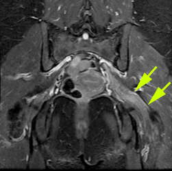
Coronal MR of sciatic nerve tumor

At our spine imaging center, leaders in neuroradiology use the most advanced equipment to perform the latest spine magnetic resonance and computed tomography diagnostic imaging and innovative image-guided therapeutic pain management services. 

These procedures may allow patients to start or resume physical therapy, more comfortably wait for their surgery date, or in some cases avoid surgery altogether.

CT-Guided Procedures

Interventional radiology techniques are used to diagnose and treat vertebral disc problems and many other conditions of the spine. Imaging technology precisely guides minimally invasive procedures with needles, catheters and other devices to alleviate back pain. Most centers offer only guidance using fluoroscopic X-rays without showing the soft tissue anatomy. At the UCSF Precision Spine and Peripheral Nerve Center, we use real time computed tomography (CT) to guide our spine injections and procedures. With CT, we can precisely deliver spinal injections while visualizing anatomy in three dimensions to maximize effectiveness and patient safety.

Our two 64 slice CT scanners are equipped with the latest radiation dose reduction software (Adaptive Statistical Iterative Reconstruction or “ASIR”) and “Smart mA,” which combine to reduce radiation dose and improve the quality of images. Together, they allow from 50 to 80 percent reduction in patient radiation dose.

Fluoroscopically-Guided Procedures

The UCSF Precision Spine and Peripheral Nerve Center offers traditional fluoroscopically-guided diagnostic spine procedures, such as myelography, discography, and lumbar puncture. Additionally, our fluoroscopy system can be used simultaneously with our CT scanner in an integrated technique that allows both visualization of complex anatomy and real-time interventions. The technique is very effective and safe for procedures such as tumor ablation, vertebroplasty , kyphoplasty, and sacroplasty.

Magnetic Resonance Procedures

Using state-of-the-art 1.5T and 3.0T MRI scanners, the UCSF Precision Spine and Peripheral Nerve Center provides the best diagnostic magnetic resonance imaging available of the spine, brain, pelvis and extremities. We offer an advanced technique, magnetic resonance (MR) neurography, which is useful for diagnosing disorders of the peripheral nerves beyond the spinal canal.

CT- guided procedures may be particularly helpful for patients who:

  • Have had extensive surgery with hardware that obscures or distorts the usual spinal anatomy.
  • Have significant osteoarthritis that may hinder access to nerves and other spinal structures.
  • Have soft tissue abnormalities such as disc herniations, cysts or scarring from prior surgery that are not visualized using conventional fluoroscopy-guided techniques.

The interventional spine techniques include:

Cisternography

In contrast to CT myelography, where the goal is to visualize structures in the spine and around the spinal cord, CT cisternography aims to study structures in and around the brain. The technique can be used, for example, to determine the location of spinal fluid leakage or to study how spinal fluid moves around the brain. Like CT myelography, CT cisternography involves injecting X-ray contrast into the fluid-filled space that surrounds the spinal cord. With this technique, contrast is usually administered by means of a lumbar puncture (also known as a spinal tap).

Discography

A discogram is a diagnostic procedure that attempts to pinpoint the cause of pain that arises from the spine. During the procedure a neuroradiologist injects contrast material into the soft/spongy discs (intervertebral disc space) that lie between the bones of the spine and X-rays and CT scans are taken of the spine following this injection.  Injecting contrast material into this area enables radiologists to pinpoint where and why there is back pain. 

Epidural Blood Patch

A unique postural headache syndrome can be seen in some patients and is caused by leakage of spinal fluid from its normal location around the spinal cord into the surrounding soft tissue structures. Spinal fluid leaks are most often spontaneous because of a ruptured perineural cyst, a disc herniation that punctures the spinal dural covering, or a result from a prior spine surgery, epidural catheterization or lumbar puncture. The leak of spinal fluid can result in abnormally low spinal fluid pressure, which allows the brain to “sag” from its normal position inside the skull, causing headaches in the upright position that are relieved in the supine position. Small leaks can be “patched” by injecting the patient's own blood around the leak through a needle inserted though the skin by our expert neuroradiologists under the careful guidance of CT. Our doctors are also experts at making the diagnosis of spinal fluid leak through the use of diagnostic MRI, CT myelography and CT cisternography. By combined use of these techniques and CT guidance, the epidural blood patch can be placed exactly at the site of the spinal fluid leak.

Epidural Injections

Epidural injections are performed to administer anti-inflammatory medication into the epidural space, the area that surrounds the spinal cord and spinal nerves. The medication reduces inflammation of nerves in the epidural space, which may reduce back pain and other symptoms caused by irritation of the spinal nerves such as numbness and tingling. A needle is inserted through the skin and deep tissues, between the bones of the spinal column, and into the epidural space using imaging guidance. The procedure can serve as an adjunct to, or in some cases, an alternative to surgery.

Facet Cyst Aspirations and Drainage

Cysts that arise from synovial disease of a facet joint may compress the spinal cord or spinal nerves and give rise to back pain or other symptoms. CT-guided cyst aspiration and drainage is a minimally-invasive treatment technique that allows temporary or permanent improvement in symptoms. Under CT guidance, a needle is inserted through the skin, soft tissues, and between the bones of the spine into the substance of the cyst. Aspirating the cyst contents results in a decrease in the size of the cyst, which reduces compression on nervous structures. When the cause of a cyst is not known, aspiration allows for a diagnosis to be made from the tissue that is removed.

Kyphoplasty, Vertebroplasty and Sacroplasty

Kyphoplasty, vertebroplasty and sacroplasty are minimally invasive procedures for treating pain from compression fractures in the bones that make up the spinal column.

In performing these procedures, our neuroradiologists use both CT and fluoroscopic image guidance to inject cement through a hollow needle into the fractured bone. The optimal pathway for placement of needles into the fracture spine is first mapped using CT and subsequently monitored with X-ray fluoroscopy. This provides the optimal three-dimensional and real-time image guidance, thus making the procedure safer and more precise.

With kyphoplasty, a balloon is first inserted through the needle into the fractured bone to create a cavity that contains the cement and prevents it from leaking into surrounding tissue. Vertebroplasty and sacroplasty are performed without the need for balloons, depending on patient anatomy. These procedures are performed under conscious sedation, so that the patient has maximal comfort, monitored by our group of advanced cardiac trained nursing staff.

Lumbar Puncture With X-Ray Guidance

A lumbar puncture is a diagnostic procedure that is performed in order to collect a sample of cerebrospinal fluid for laboratory analysis. The study is performed using X-ray fluoroscopy, or in rare instances CT scanning.

Myelography

Myelography is an imaging examination in which X-ray contrast is injected into the space around the spinal cord (the subarachnoid space) and spinal nerve roots. using a real-time form of X-ray called fluoroscopy. A CT scan of the area of interest typically is obtained following the myelogram. Your doctor may order this rather than, or in addition to, an MRI scan of the region, depending on the result of the MRI scan or what kind of abnormality is suspected. Learn more about how to prepare for a myelogram.

Nerve Block

A nerve block is a CT-guided injection of anesthetic or anti-inflammatory medication targeted toward a certain nerve or group of nerves to treat pain. The purpose of the injection is to either determine the location of the pain generator or decrease inflammation, and to "turn off" a pain signal coming from a specific location in the body.

The neuroradiologist places the needle in exactly the right location using CT scanning (CAT scans). This "imaging guidance" allows the patient to receive maximum benefit from the injection.

You can obtain more information on Nerve Block from the RadiologyInfo website.

Radiofrequency Ablation

Radiofrequency Ablation (RFA) is a method used to treat certain tumors, such as osteoid osteoma (a benign tumor of the bone), or the sensory nerves that supply painful areas of the back, such as facet joints. During this procedure the tumor or the nerve is heated by the application of radiofrequency energy via a special needle placed precisely in the area of interest for a period of approximately 6 minutes. This procedure is minimally invasive, is done on an outpatient basis, and has a short recovery time. Patients are candidates for RFA treatment if they have shown pain relief from medication nerve blocks.

Spinal Biopsy

Spinal biopsy under imaging guidance is often necessary to establish a diagnosis of infection or benign and malignant tumors. In these cases, imaging is absolutely essential to identify the safest access to the most suspicious areas of abnormal tissue or bone. Unlike fluoroscopy, CT imaging guidance maximizes the diagnostic yield of a biopsy by identifying the sites most likely to be abnormal and allows safe percutaneous access to the biopsy site by avoidance of critical structures around the spine such as large blood vessels, nerves, and the spinal cord. Biopsies can be performed with a small (fine needle aspiration) or large (core biopsy) needle technique depending on the clinical indication.

Spinal Tumor Ablation

Spinal tumor ablation is a new approach for improving the safety profile of vertebroplasty and kyphoplasty in cases of vertebral compression fractures that are caused by malignant tumors. Many cancers such as breast cancer, melanoma, renal carcinoma and multiple myeloma frequently involve the bones of the spine and lead to painful fractures. In cases where a tumor has destroyed the vertebral body, kyphoplasty and vertebroplasty cannot be performed safely. Spinal tumor ablation techniques utilize CT guidance to remove portions of the tumor through a needle inserted through the skin. While this is not effective treatment for the tumor itself, it does allow creation of a cavity in the tumor through which kyphoplasty or vertebroplasty can be safely performed to treat spinal pain.

Sympathetic Ganglion Block

In addition to selective nerve block, using three-dimensional imaging guidance it is also possible to selectively block sympathetic ganglia. For example, our neuroradiologists have performed blocks of the stellate and impar ganglia for treatment of medically intractable complex regional pain syndrome and pain from certain sacral disorders, respectively.