Current and Future Applications of 3D Printing in Breast Cancer Management
3D printing technology has been around for about 40 years. In medicine, we have seen rapid expansion across almost every subspecialty from pre-surgical planning to production of patient-specific surgical devices to simulation and training. However, there is a notable lack of literature on the applications of 3D printing in breast cancer management.
Tatiana Kelil, MD is an assistant professor of clinical radiology in the Breast Imaging section at the UC San Francisco Department of Radiology and Biomedical Imaging and serves as a co-director for the UCSF Center for Advanced 3D+ Technologies (CA3D+). Dr. Kelil is interested in advanced imaging visualization techniques and has experience with 3D post-processing, patient-specific modeling, and medical 3D printing. Dr. Kelil, along with Arpine Galstyan (MD candidate at UCSF) were corresponding authors on a review and discussion of the current applications of 3D printing in breast cancer management and potential impact on future practices. The paper was recently published in 3D Printing in Medicine.

Their review looked closely at five different applications of 3D printing in breast cancer management:
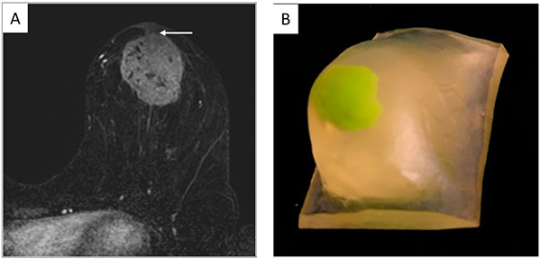
- Breast-conserving surgery (BCS) and tumor localization
- Breast reconstruction surgery
- Physician-patient and interdisciplinary communication
- Education and simulation
- Quality control
“Overall, 3D printing is fundamentally changing breast cancer surgery. It allows for patient-specific presurgical planning and more customized surgical intraoperative surgical guides for both breast conservation and breast reconstruction surgeries,” says Dr. Kelil.

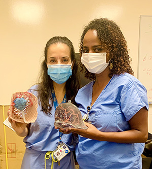
This review was made possible through a collaborative team effort. Michael Bunker from the UCSF CA3D+ participated in the creation of 3D printed models along with Fluvio Lobo, Robert Sims, James Inziello and Jack Stubbs from the University of Central Florida. Surgical procedures were performed by Rita Mukthar, MD from the UCSF Department of Surgery.
 “Dr. Mukthar and I collaborate often to create precise 3D model of patients’ breasts for cancer surgeries,” says Dr. Kelil. “In addition to leading to better aesthetic surgical outcomes and negative surgical margins, these models serve as teaching and explanation tools for patients and trainees, and they can improve interdisciplinary communication between healthcare providers.”
Here at UC San Francisco, the CA3D+ was formed as a multidisciplinary collaboration between the departments of Pediatric Cardiology, Orthopedics and Radiology and Biomedical Imaging thanks to a grant awarded by the School of Medicine. The CA3D+ uses 3D printing and virtual reality to help doctors prepare for surgery and explain procedures to patients.
According to the team, emerging fields for 3D printing and breast cancer management include bioprinting and personalized radiation therapy to address challenges encountered with current breast cancer management approaches.
