Instrumentation and Physics Research
Instrumentation and physics research in our lab is focused on advancing our understanding of hardware and software approaches to quantitative molecular imaging.
Current Research Highlights
Dynamic cardiac SPECT and PET: We are developing, implementing, and utilizing dynamic cardiac SPECT and PET imaging methods that provide quantitative metrics like myocardial blood flow and coronary flow reserve for studying cardiac diseases. Funding support: NIH/NHLBI (R01HL135490, R01HL050663), DOE, etc. |
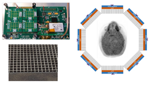
Variable aperture, energy-indepedent CZT-SPECT: We are developing a next-generation radiation detection technology for SPECT imaging using solid-state direct-coversion radiation detector, CZT, a novel ASIC, and a novel design of radiation collimator that can be used for a wide spectrum of gamma-emitting radionuclides without needing to change collimators between studies. Funding support: NIH/NIBIB (R01EB026331, R01EB012965), etc. |
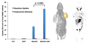
Theranostic molecular imaging probe and application: We help develop novel theranostic molecular probes such as 124I-MIBG for 131I-MIBG therapy and other beta- and alpha-emitting thepeutic radiopharmaceuticals, and are developing robust and accurate radiation dosimetry methods for tumor and normal tissues. Our particular research interests are, but not limited to, neuroblastoma and prostate cancer. Funding support: NIH/NCI (R01CA154561), V Foundation, Alex's Lemonade Stand Foundation, etc. |
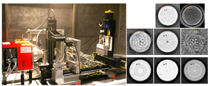
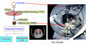
X-ray dark-field/phase-contrast imaging technology: We are developing a next-generation x-ray phase-contrast/dark-field imaging technology. A few different technologies are being evaluated, and our efforts are in close ties with industry efforts in this area. Funding support: NIH/NIBIB (R43EB027535), etc. |
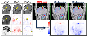
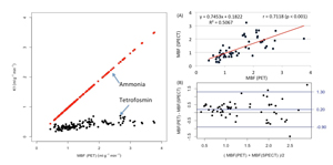
Quantitative PET/MRI, PET/CT, and SPECT/CT: We are developing techniques to improve quantitative accuracy of PET reconstruction in PET/MRI and PET/CT and SPECT reconstruction in SPECT/CT by implementing new methods of attenuation, collimator response, partial volume, and motion corrections using modern computational approaches like compressed sensing, deep neural networks, etc. Funding support: NIH/NCI (K25CA114254, R21CA086893), University of California, Siemens, GE Healthcare, etc. |
Past Research Highlights
Simultaneous Emission-Transmission CT (ETCT): UCSF PRL, led by the late Professor Bruce Hasegawa, developed a new technology called, simultaneous emission-transmission computed tomography (ETCT), a predecessor to dual-modality SPECT/CT. Selected publication(s): Hasegawa BH, et al. Description of a simultaneous emission-transmission CT system. SPIE Vol 1231 Medical Imaging IV Image Formation (1990). |
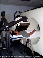
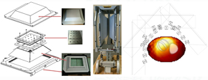
Clinical SPECT/CT prototype: UCSF SPECT/CT system (left figure) was built in 1995, with two imaging modalities (CT and SPECT) sharing a common patient table. This was the first clinical dual-modality functional-anatomical imaging system that was actually built and tested. |
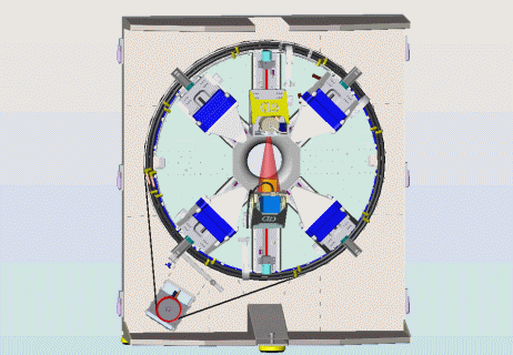
'MoHawk' Small Animal SPECT/CT: This is a system of two tungsten pinhole CZT-based gamma cameras and x-ray source and detector built on a mechanical platform powered by a slip ring. Many members of UCSF PRL, under supervision of the late Professor Bruce Hasegawa, have worked on this innovative imaging system. Funding support: NIH/NIBIB (R01EB000348), University of California, Gamma Medica, etc. Selected publication(s): Izaguirre EW, et al. Imaging structure and function in small animals. Presented at the sixth Bioengineering Research Partnership Grantee Meeting, North Bethesda, MD, 2006. |
Multipinhole SPECT for heart and brain imaging: Our laboratory has simulated, designed, and fabricated a 20-pinhole collimator that fits with conventional gamma camaras for high-sensitivity and high-resolution heart and brain imaging. Funding support: NIH/NHLBI (R21HL083073), GE Healthcare, etc. |