Tijana Popovic, MS Spotlight

February 27, 2023
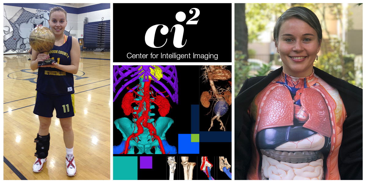
Tijana Popovic, MS, a Clinical Imaging Analyst at the 3DLab within UCSF’s Center for Intelligent Imaging (ci2), has pursued avenues to enhance human ability through fitness and science. This emerges through her commitment to both athletics and mental wellness, and her work creating 3D imaging models that augment radiologists’ ability to see inside patients’ bodies with an accuracy and holistic vision beyond traditional imaging slices.
Tijana Popovic grew up in the city of Nis, Serbia where she understood the value of being a member of a high-level team long before she came to San Francisco to be a player on our Team Science. While studying full time at college, Tijana was also a professional basketball player, representing Nis in the national pro-basketball league for six years.
Reflecting on how her background in competitive team-sports shaped her approach to science and medicine, Tijana observed, “Every game and every new encounter is a challenge. You need to prepare and perform your part well, while always being open to shift tactics as you go.”
Tijana’s lifelong fascination with the human body and what allows us to perform the everyday feats that most of us take for granted drew her to the medical field. Her experience in professional sports gave her first-hand experience with the potentially serious injuries that arise from pushing a body to perform at elite levels. Her career path demonstrates her desire to enhance our ability to know what is happening inside the body utilizing the tools of biomechanical research or clinical imaging.
As Tijana says, “I got into medical science because the human body is amazing. There are a lot of injuries that I experienced or that people around me experienced, and for some it is a life-changing moment while others go back to running ten days after a knee surgery. After putting so much stress on my body earlier in life I still run, play soccer, and spend all the time I can outside and stay active. I am fascinated by the mechanics and forces behind this.”
Popovic joined UCSF in 2016 to research biomechanics. In the Department of Radiology and Biomedical Imaging, in addition to imaging, she researched the mechanical side of movement with motion tracking to gather information about the way people move, going beyond human senses to use latest motion capture technology, including stick-on trackers, and cameras to create an observation system akin to how Hollywood movies might capture an actor’s performance for CGI. Tijana said, “The way we move has such a great impact on our quality of life at an everyday level. I was drawn to trying to figure out how biomechanics (forces, joint angles, moments, etc.) relate to hip joint health.”
Tijana places great value in UCSF’s strong culture of mentorship, saying “I am very grateful for all the great mentors and supervisors I had here. My superstar mentor in biomechanics was Richard Souza, PT, PhD. He didn’t tell you what to do, he will lead us to what to do. He leads with support from behind the scenes, not just with personality from the front.”
After a few years in biomechanical research, Tijana chose to make the transition to the medical side of treatment, entering Imaging as part of the 3DLab at ci2. The 3DLab, based at China Basin, operates as a service available to surgeons and doctors throughout UCSF Health with the goal of improving the results of conventional image reads by rendering them in 3-D models. The lab augments the ability of human image readers by enhancing traditional 2-D slices into 3-D models with the assistance of AI which cleans up image noise. This process facilitates the interpretation of scans and grants the human image reader the ability to spot features that would otherwise be hidden and escape notice. Tijana appreciates that this work provides both the intellectual allure of cutting-edge medical science in a wide variety of cases and the satisfaction of a direct and concrete role in improving patient outcomes.
Tijana finds fulfillment in the 3DLab’s direct connection to patient health, saying, “I really enjoy my work because I know I am helping people. In biomechanics we were also giving insight about what was happening inside the body, but with imaging you actually can see into the tissue at the cellular level. Imaging has given me another perspective on what I was already interested in. Going to work is like meditation for me, it improves my mental wellbeing.”
Tijana creates 3-D reconstructions of the human body that can help surgeons prepare for surgery and assists doctors in performing the measurements necessary to assess recovery and plan the next steps. She described how these 3-D reconstructions are a key part of enhancing the ability of human clinicians and researchers through the tools of AI and other advanced systems, “A spatial representation of the human body can open your eyes in a way not otherwise possible. With this, surgeons can see inside to improve surgical outcomes, especially in very complex situations. Imaging, even more than biomechanics, allows us to see inside to the tissue or even cellular level and give a new perspective of what is actually happening. When you can see the problem better, the outcome for patients improves.”
The 3DLab at ci2 may have access to the cutting edge of AI, but the work Tijana undertakes with the 3DLab is no simple matter of directing the advanced programs at the input and then waiting for them to spit out a result. Tijana joked, “Before I entered Imaging, I imagined that the AI program was doing everything for you and also making all of the measurements, and you just had to kind of click and check. That is not true at all.” Tijana and her colleagues choose exactly how to apply the programs and how to guide interpretation of the raw output into a visual useful for human eyes, which is a delicate art.
Further, the clinical imaging analyst must decide the exact measurements to pursue, what type of 3-D reconstruction is appropriate for the case, and what protocols should be used in the case of congenital malformations or other patient variations. There is a lot of work that gets done behind the scenes in preparing a 3-D model, particularly when it comes to cleaning up imaging noise. An analyst needs to know the anatomy and be able to understand the potential variations. Cases generally come to the lab because they are complex, and in those cases the standard rules often do not apply. “Here at 3DLab we have standard procedures, but we are always open to change when it will produce more successful results. You have to be fast on your feet and quick to shift tactics.”
Tijana provided the example of a hypothetical musculoskeletal image sent to the lab to develop a model image of a bone fracture. If the patient has had a hip replacement or a knee replacement, that hardware will throw a tremendous amount of noise into the imaging. It takes a practiced and knowledgeable eye to help the image processing system understand what is bone and what is noise.
 Beyond the lab, Tijana is a mother to a two-year-old daughter and loves spending time outside, always moving. Tijana is grateful that California has given her the opportunities for such activities, as she believes mental health improvement is as important as physical health. Like many others, she has come to miss talking to people as easily since the beginning of remote work. A social extrovert, Tijana is not just one to make friends at events, meet-ups, or at the gym, but is very used to meeting people on the street, BART, and anywhere else she intersects with humanity.
“We’re all so different in this country which is so great. You can hear a lot of wonderful stories and learn from them. The best advice I was ever given is; be curious about things. Everyone can teach you something, you just need to listen. And then be open to change.”