Celebrating 20 Years of Innovation: UCSF’s Center for Molecular and Functional Imaging
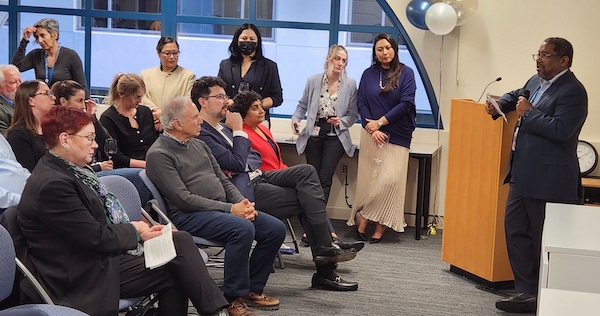
Twenty years ago, the Center for Molecular and Functional Imaging (CMFI) at China Basin Landing opened its doors with San Francisco’s first 3T MRI and two 1.5T clinical scanners, followed a few years later by a cyclotron which launched our in-house radiopharmaceutical program. Since then, the CMFI has been at the forefront of 3T research and major advances in theranostics and precision medicine. We’re pleased to share highlights and milestones achieved along the way to this 20th anniversary and look forward to celebrating the CMFI’s first two decades with a reception in January.

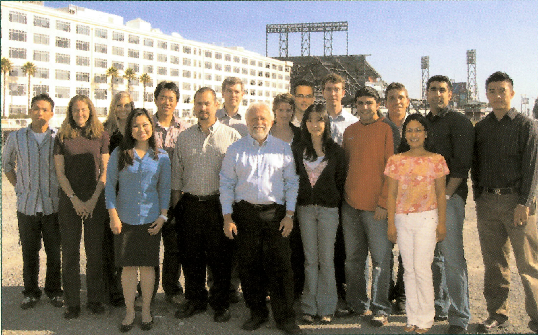
In the early 2000s, Ronald Arenson, MD, chair of UCSF’s Radiology Department from 1992 to 2017, knew the department needed to expand. “We were totally out of space for research and clinical work at Parnassus. An external review of on-campus construction predicted a very high cost-per-square-foot, but at the moment San Francisco was in the aftermath of the dot-com bust with plummeting commercial real-estate prices. One of our vendor partners mathematically evaluated the optimum location in the city for clinical space, and on that map there was a bull’s-eye over China Basin.”
The CMFI space was built on time and within budget on the third floor of the China Basin Landing and on December 12, 2003, opened with two research instruments: the pioneering 3T MRI and a 16-slice CT. Sharmila Majumdar, PhD, explained the impact of this new technology, “It was the first 3T magnet in San Francisco, and one of the first in any public institution. This made UCSF a leader in high-field imaging.” Dan Vigneron, PhD, also reflects on those early days of high field 3T MRI, “I received a grant to develop unprecedented 3T prostate cancer MRI at a time when 3T was thought to only be useful for brain imaging. But now due to the pioneering work at China Basin, 3T is now state-of-the-art for prostate imaging and other body applications. Also, new high-sensitivity multichannel brain coils were developed in the MRI electronics shop there, and these types of detectors are now widely used worldwide.”
The China Basin facility provided a home for radiology faculty representing physics, nuclear medicine, bioengineering, musculoskeletal research, and neuroradiology. UCSF Health also opened an outpatient imaging location in the same building, and the co-location of these powerful research scanners fostered immediate benefits and a seamless flow between clinical needs and CMFI’s research.
Youngho Seo, PhD, who, with Bruce Hasegawa, PhD, was one of the first researchers to move his lab into CMFI, described the culture that grew there, “There is power in a close network of researchers. The UCSF approach is to bring in many individual faculty who can establish themselves and their work, rather than relying on one figurehead researcher.” Arenson agreed with this assessment and the results it produced, and noted that “After we opened China Basin, UCSF rose up to be number two or three in the nation in federal (NIH) funded research. The 3T MRI was pioneering in research and it is now UCSF’s clinical norm.” This reflects the rapid translation of research into clinical arenas for which the department is renowned.
The CMFI’s 3T research program generated UCSF’s first 3T prostate exams, first 3T knee images, cutting-edge brain tractography research, and the diffusion imaging program that now is a cornerstone in neurosurgery clinical evaluations. The Center’s first external research collaboration was a project between UCSF professor Ben Francs, MD, MS, MBA, and Clifford Berkman, PhD, of San Francisco State University, who developed early prostate-specific membrane antigens (PSMAs)-targeting PET and SPECT agents for prostate cancer imaging. Now, faculty members Michael Evans, PhD, and Robert Flavell, MD, PhD, lead research on novel theranostic compounds which use paired isotopes to combine imaging and therapy in one molecular structure, and which are in the process phased human trials toward potential FDA approval. Every year, the research group submits two to three Investigational New Drug (IND) applications to the FDA for new compounds submitted for human trials. Henry VanBrocklin, PhD, director of the Radiopharmaceutical Research Program within CMFI, described this pace of constant science, “We’ve translated a number of new radiopharmaceuticals here and have seen significant growth of our radiopharmaceutical research program.”
CMFI’s early days were marked by prominent investments in growth and infrastructure. On Labor Day 2005, the cyclotron was installed in the basement of CMFI’s building, and by December of 2006, UCSF produced its first FDA-approved clinical radiopharmaceutical. Seo described how the cyclotron has facilitated CMFI’s growth, “We are very good at first-in-human imaging studies; they are our forte. We know how to develop compounds from the bench and on into human trials.” Now, with theranostics and precision medicine, CMFI’s close linkage between research and clinical implementation keeps them at the forefront of this rapidly expanding field.
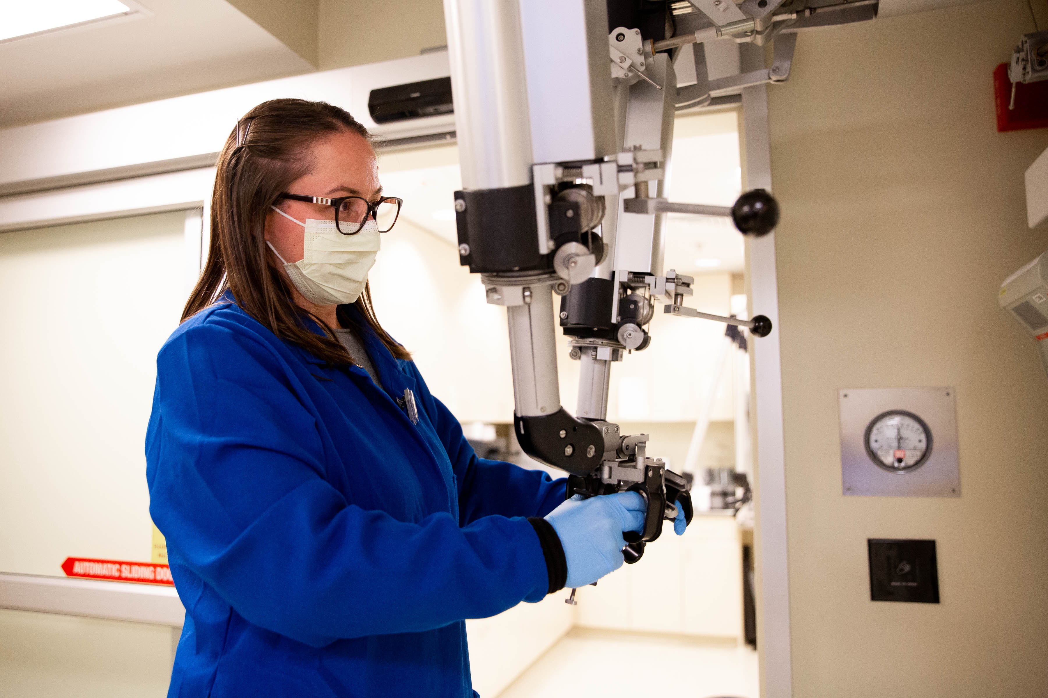
An FDA registered cyclotron both expanded UCSF’s capability for clinical applications and gave access to new radiopharmaceuticals for research. Gone were the days of driving compounds through traffic across the Bay Bridge from Berkeley, and now UCSF researchers are developing many new radiopharmaceutical with potential for translation for human imaging and therapy. VanBrocklin has developed over 10 compounds with FDA approval, and has provided leadership to help vital work like Seo’s research on radionuclide and x-ray imaging instrumentation and physics, Evans and Flavell’s theranostics breakthroughs, and Dave Wilson, MD, PhD’s development of new molecular probes for infection and brain imaging, as well as new agents for hyperpolarized 13C spectroscopy with positron emission tomography (PET) correlates for imaging.
Today, Seo, from his desk in the office that formerly seated the late medical physicist Bruce Hasegawa, PhD, is proud of what he and his CMFI colleagues have accomplished in 20 years, “Our people are very successful today and all help CMFI do great things.”


